Research
When CNNs Outperform Transformers and Mambas: Revisiting Deep Architectures for Dental Caries Segmentation
When CNNs Outperform Transformers and Mambas: Revisiting Deep Architectures for Dental Caries Segmentation Overview Research area: Medical image segmentation — specifically automated pixel-level segme
- arXiv
- 2511.14860
- Published
- 2025-11-18
- Authors
- Aashish Ghimire, Jun Zeng, Roshan Paudel, Nikhil Kumar Tomar, Deepak Ranjan Nayak, Harshith Reddy Nalla, Vivek Jha, Glenda Reynolds, Debesh Jha
AI summary
When CNNs Outperform Transformers and Mambas: Revisiting Deep Architectures for Dental Caries SegmentationOverview
Research area: Medical image segmentation — specifically automated pixel-level segmentation of dental caries (tooth decay) in panoramic radiographs, spanning convolutional neural networks (CNNs), vision transformers, and state-space "Mamba" architectures.
Technical level: Intermediate. The paper assumes familiarity with encoder–decoder segmentation networks, attention mechanisms, and standard segmentation metrics, but its central argument (architecture-task alignment matters more than architectural novelty) is accessible to a general machine learning audience.
Scope: A single-dataset, unified-protocol benchmark of 12 segmentation architectures evaluated on the DC1000 panoramic radiograph dataset, comparing CNNs against transformer- and Mamba-based models on accuracy, boundary quality, and computational efficiency.
What This Paper Is About
Automated detection of dental caries in panoramic X-rays is difficult because lesions have low contrast against surrounding tissue, vary in shape and location, and annotated data is scarce. The paper asks whether the field's recent shift toward complex attention-based and state-space architectures actually improves segmentation performance for this specific domain, or whether simpler CNNs remain better suited. To answer this, the authors train 12 state-of-the-art segmentation models under one identical pipeline on the DC1000 dataset and compare them head to head.
Key Contributions
-
First comprehensive cross-architectural benchmark for dental caries segmentation. The paper evaluates 12 segmentation architectures — spanning CNNs (U-Net, DoubleU-Net, ResUNet++, ColonSegNet), transformers (TransNetR, TransRUPNet, RSAFormer, PVTFormer), and Mamba/state-space models (VMUNet, VMUNetV2, MambaUNet, RMAMamba-S) — on the DC1000 panoramic radiograph dataset, all trained and evaluated under identical data splits, augmentations, and optimization settings.
-
Evidence that CNNs dominate on this task. The top three results across all reported performance metrics were achieved by CNN-based architectures. DoubleU-Net reached the highest mDSC of 0.7345, mIoU of 0.5978, and Precision of 0.8145, while the best transformer (PVTFormer, mDSC 0.6733) and the best Mamba model (RMAMamba-S, mDSC 0.6583) fell short. The paper reports this as roughly 9% higher mDSC achieved with significantly fewer parameters and faster inference.
-
Joint accuracy-and-efficiency analysis, including a discrimination/segmentation mismatch. Beyond overlap metrics, the authors report GFLOPs, parameter counts, and FPS for every model, plus ROC analysis showing that Mamba and transformer models can attain higher AUC (MambaUNet reached the overall highest AUC of 0.9816) while still producing worse segmentation masks — illustrating that pixel-level discriminability does not guarantee anatomically meaningful boundaries.
-
Clinical framing and a released codebase. The paper connects results to computer-aided dental diagnosis, discussing where each architecture family succeeds and fails qualitatively, and releases code at https://github.com/JunZengz/dental-caries-segmentation.
Main Findings
-
DoubleU-Net is the strongest overall model. It achieved the best mIoU (0.5978), mDSC (0.7345), and Precision (0.8145), with Recall 0.7009, F2 0.7115, and Hausdorff Distance (HD) 2.0260. The authors attribute this to its stacked dual-decoder design for capturing minute carious regions.
-
CNN-based models take the top three slots on every metric. After DoubleU-Net came U-Net (mIoU 0.5836, mDSC 0.7236, Recall 0.7159, Precision 0.7649, F2 0.7161) and ColonSegNet (mIoU 0.5669, mDSC 0.7044, Recall 0.6819, Precision 0.7779, F2 0.6877).
-
U-Net has the lowest boundary error. U-Net reported the lowest HD at 2.0022, followed by DoubleU-Net at 2.0260 and ColonSegNet at 2.0686. The authors note this balance makes U-Net favorable for real-time or near-real-time diagnostic settings.
-
Transformers cluster below CNNs. PVTFormer led the transformer family (mIoU 0.5291, mDSC 0.6733, Recall 0.6564, Precision 0.7461, F2 0.6594, HD 2.1637), closely followed by TransRUPNet (mIoU 0.5278, mDSC 0.6723). RSAFormer was weakest among transformers (mIoU 0.4465, mDSC 0.5916) and had the highest HD of any model at 2.3834.
-
Mamba models are efficient but imprecise. RMAMamba-S was the best Mamba model (mIoU 0.5124, mDSC 0.6583, Recall 0.6366, Precision 0.7348, F2 0.6424, HD 2.2225). VMUNetV2 was the weakest overall model (mIoU 0.4039, mDSC 0.5549, Recall 0.5516, Precision 0.6308, F2 0.5475, HD 2.3620), and MambaUNet reported an HD of 2.3653.
-
Efficiency favors CNNs at the inference level. U-Net was fastest at 40.83 FPS, with ResUNet++ second at 34.22 FPS and DoubleU-Net third at 33.09 FPS. The slowest model was RSAFormer at 9.95 FPS, and RMAMamba-S ran at 11.43 FPS. ResUNet++ had the fewest parameters (4.06M), followed by ColonSegNet (5.01M) and MambaUNet (15.48M).
-
Mamba models have the lowest GFLOPs. VMUNet reported 9.25 GFLOPs and VMUNetV2 9.90 GFLOPs, versus 147.43 for U-Net, 139.86 for ColonSegNet, and 97.64 for PVTFormer. RMAMamba-S (27.95 GFLOPs, 55.21M parameters) and RSAFormer (31.69 GFLOPs, 65.76M parameters) were the heavyweights in their families.
-
High AUC does not translate into good masks. All models exceeded AUC > 0.87. Among CNNs, DoubleU-Net scored the highest AUC at 0.9467; among transformers, PVTFormer reached 0.9269. Mamba models scored highest on discrimination — VMUNet and VMUNetV2 both at 0.9782, and MambaUNet at the overall highest 0.9816 — yet produced the weakest overlap and boundary metrics, often over-segmenting and missing lesion contours.
-
Qualitative behavior differs by family. In the visual comparison (Figure 3), DoubleU-Net preserved subtle radiolucent boundaries and diffuse enamel–dentin transitions; U-Net showed strong structural localization but under-segmented low-contrast regions; PVTFormer captured broader lesion context but over-segmented in response to intensity variation and metallic artifacts; RMAMamba-S produced smoother, less precise boundaries and missed fine lesion detail.
-
Sparsity of lesions amplifies error. Because carious regions occupy only a fraction of each radiograph, missing pixels disproportionately affect mIoU and Dice, which the authors cite as a partial explanation for recall-versus-precision differences and for the sensitivity of boundary measures like HD.
Methodology in Plain English
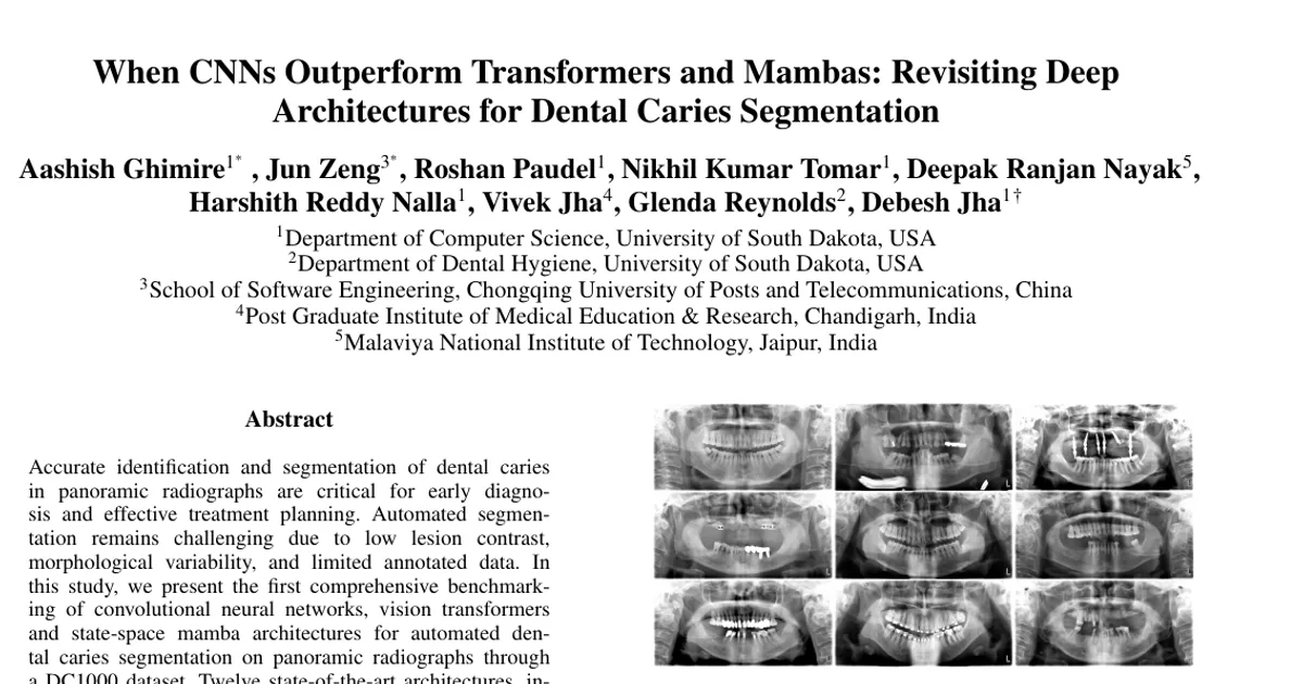
Data. The study uses the DC1000 dataset: 597 high-resolution panoramic radiographs with pixel-level annotations of dental caries made by experienced dentists. Of these, 497 are training images (4 of which lack masks) and 100 are test images. The dataset provides no official validation split, so the authors optionally split the training set 80:20 for training and validation. Images are stored as 8-bit grayscale, with binary masks where 0 is background and 1 is a carious lesion.
Fair comparison. All 12 models were trained on the PyTorch framework on a single NVIDIA V100 GPU with 32GB memory, using identical data splits, augmentations, and optimization settings. Details: Adam optimizer; a combined loss of binary cross-entropy with logits (pos_weight = 18.0) and focal loss (alpha = 0.75, gamma = 2.0) mixed at a 0.5:0.5 ratio; learning rate of 1×10⁻⁴ with a ReduceLROnPlateau scheduler (patience = 5); 500 epochs with early stopping on validation Dice (patience = 50); batch size 4; and input size 384×384 as 3-channel RGB panoramics.
Augmentation and preprocessing. Training images were resized to 384×384, randomly horizontally flipped (p = 0.5), shifted (±5%), scaled (±5%), rotated (±15°, border = constant, p = 0.5), and given random brightness and contrast adjustment (p = 0.3). Contrast-limited adaptive histogram equalization was also included among the augmentations. Validation images were only resized and normalized. The best checkpoint was selected by highest validation IoU to prevent overfitting.
Evaluation. Models were compared on mean Intersection over Union (mIoU), Dice coefficient (mDSC), Recall, Precision, F2-score, and Hausdorff Distance (HD), plus GFLOPs, parameter count, FPS, and ROC/AUC. Qualitative overlays were inspected, and figures were plotted to visualize convergence trends across architectures.
Reference comparison. The paper also tabulates prior dental deep learning studies across modalities (intraoral photographs, panoramic radiographs), including work by Park et al. (2022, 2,348 images from 445 patients), Zhu et al. (2022, 1,159 images with 3,217 annotated caries lesions), Asci et al. (2024, 6,075 pediatric images), Hao et al. (2024, TSI15k with 1,589 labeled and 14,728 unlabeled images), Lim et al. (2025, 597 images split into 443 train / 50 val / 100 test), and Pornprasertsuk-Damrongsri et al. (2025, 500 images with 14,997 teeth).
Why This Matters
Impact on research. The paper pushes back on the assumption that architectural complexity automatically improves medical image segmentation. It argues that architecture–task alignment, spatial inductive priors, data scale, and the ability to capture fine-grained texture cues matter more than novelty, and it provides a standardized 12-model baseline where the field previously relied on differing protocols and non-comparable results.
Real-world applications:
- Computer-aided dental diagnosis that flags carious lesions on routine panoramic X-rays, supporting earlier treatment planning.
- Screening in settings with limited specialist availability, where a fast automatic pre-read (U-Net ran at 40.83 FPS and DoubleU-Net at 33.09 FPS) could triage which radiographs need expert review.
- Lesion severity and treatment-need assessment, which depends on accurate boundary localization — the area where DoubleU-Net performed best.
- Integration into dental practice software, where parameter and compute budgets are constrained; ResUNet++ (4.06M parameters) and MambaUNet (15.48M parameters) show the possible low-cost end of the spectrum.
Industry relevance. Any organization deploying dental AI must choose among models with very different cost profiles — from 9.25 GFLOPs for VMUNet to 147.43 GFLOPs for U-Net — and the paper shows the highest-AUC model is not the best segmenter. That disconnect matters for vendors, regulators, and clinicians evaluating claims based on discrimination metrics alone.
Future Directions
- Build larger, multi-institutional datasets. The authors identify the relatively small dataset size as a likely reason transformers and Mamba models underperformed, and propose multi-institutional data collection as future work.
- Extend to more dental classes. The paper suggests expanding beyond caries to conditions such as impacted tooth and periapical radiolucency.
- Test on more heterogeneous cohorts. The plan includes evaluating the algorithms on more diverse patient populations than the current DC1000 cohort, which already spans full dentition, missing teeth, and orthodontic appliances.
- Address class imbalance and low-contrast lesions. The authors flag these as factors that may have limited performance for all models, leaving open how to design architectures or training schemes that handle sparse, subtle lesions with better boundary precision.
Target Audience
This paper is most useful for medical imaging researchers and graduate students benchmarking segmentation architectures; dental AI developers selecting a backbone under data and compute constraints; and clinical informatics teams or practitioners evaluating whether an attention-based model is worth adopting over a well-tuned CNN. Readers already familiar with segmentation benchmarks will find the efficiency tables and the AUC-versus-overlap discrepancy the most actionable parts.
Authors’ abstract
Accurate identification and segmentation of dental caries in panoramic radiographs are critical for early diagnosis and effective treatment planning. Automated segmentation remains challenging due to low lesion contrast, morphological variability, and limited annotated data. In this study, we present the first comprehensive benchmarking of convolutional neural networks, vision transformers and state-space mamba architectures for automated dental caries segmentation on panoramic radiographs through a DC1000 dataset. Twelve state-of-the-art architectures, including VMUnet, MambaUNet, VMUNetv2, RMAMamba-S, TransNetR, PVTFormer, DoubleU-Net, and ResUNet++, were trained under identical configurations. Results reveal that, contrary to the growing trend toward complex attention based architectures, the CNN-based DoubleU-Net achieved the highest dice coefficient of 0.7345, mIoU of 0.5978, and precision of 0.8145, outperforming all transformer and Mamba variants. In the study, the top 3 results across all performance metrics were achieved by CNN-based architectures. Here, Mamba and transformer-based methods, despite their theoretical advantage in global context modeling, underperformed due to limited data and weaker spatial priors. These findings underscore the importance of architecture-task alignment in domain-specific medical image segmentation more than model complexity. Our code is available at: https://github.com/JunZengz/dental-caries-segmentation.