Research
SAR2Net: Learning Spatially Anchored Representations for Retrieval-Guided Cross-Stain Alignment
Overview Research area: Computational pathology / medical image analysis — specifically cross-stain whole-slide image (WSI) registration and region-level alignment between HE and IHC slides. Technical
- arXiv
- 2610.26217
- Published
- 2026-10-19
- Authors
- Tianle Shen, Fang Yan, Xiaofan Zhang
AI summary
Overview
Research area: Computational pathology / medical image analysis — specifically cross-stain whole-slide image (WSI) registration and region-level alignment between HE and IHC slides. Technical level: Advanced (assumes familiarity with contrastive learning, feature retrieval, and WSI registration pipelines). Scope: The paper proposes SAR2Net, a retrieval-based framework that learns deformation-invariant "spatially anchored" point representations so that HE and IHC slides can be matched region-by-region without any global pre-alignment.
What This Paper Is About
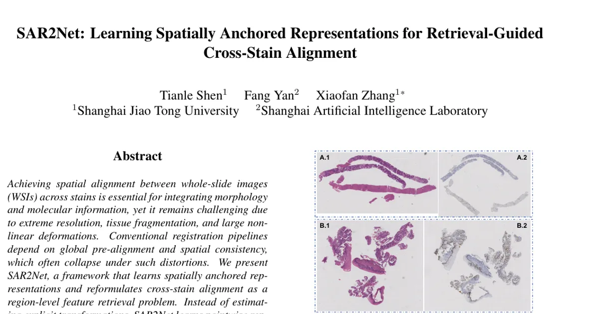
When pathologists want to compare tissue morphology (shown by HE staining) with molecular expression (shown by IHC staining), they need to know which region of one slide corresponds to which region of the other. Existing automated methods treat this as image registration: they first roughly line up the two slides globally, then refine locally. That assumption collapses for biopsy specimens, where tissue is torn, bent, fragmented, or arranged in multiple disconnected pieces, so much of one slide has no roughly-overlapping counterpart. The authors instead reframe the problem as region-level feature retrieval, where each point is described by its geometric relationship to nearby tissue landmarks rather than by image appearance or a global transformation.
Key Contributions
- SAR2Net network: A model that encodes the local geometric relations between a query point and a set of spatial anchors, producing "spatially anchored" representations that stay consistent across stains and deformations.
- Two-stage retrieval pipeline: A retrieval-guided alignment procedure that establishes region-level correspondences even when global alignment is infeasible, with a second refinement round inside each matched region.
- Anchor extraction and grouping strategy: Landmarks are extracted with pretrained SuperPoint/SuperGlue on low-resolution thumbnails, with rotation-aware merging and RANSAC-based geometric consensus to keep only locally coherent anchor subsets.
- State-of-the-art results on a biopsy-oriented dataset: 370 HE–IHC slide pairs from Xijing Hospital, where approximately 35% of slides exhibit severe tissue distortions or multiple disjoint fragments — conditions the authors note are rarely represented in existing benchmarks.
Main Findings
- Large gains over registration baselines: On the biopsy dataset, SAR2Net with second-round refinement reaches mIoU 0.899, mDice 0.942, aw-IoU 0.901 and aw-Dice 0.944, versus RegWSI at 0.691 / 0.786 / 0.699 / 0.794 and VALIS at 0.635 / 0.705 / 0.647 / 0.716. The single-round variant already reaches 0.891 / 0.938 / 0.893 / 0.939.
- Refinement helps consistently: The second round adds a further gain over the single-round variant on all four overlap metrics, with the improvement concentrated in the high-threshold ranges (IoU 0.7–0.9 and Dice 0.8–0.9).
- More uniform quality across region sizes: VALIS and RegWSI show area-weighted metrics slightly above their mean metrics, which the authors attribute to a few large, easy-to-match regions dominating; SAR2Net shows only a minimal gap between mean and area-weighted metrics, indicating consistent alignment for large and small regions alike.
- Much higher success rates: At IoU 0.75 the success rate is 0.939 for the full method versus 0.594 (VALIS) and 0.545 (RegWSI); at IoU 0.85 it is 0.812 versus 0.429 and 0.324; at Dice 0.75 it is 0.974 versus 0.690 and 0.740; at Dice 0.85 it is 0.946 versus 0.606 and 0.574.
- Random perturbations are the biggest single ablation factor: Removing them drops mIoU from 0.891 to 0.847, mDice from 0.938 to 0.911, and aw-IoU from 0.893 to 0.852. Similarity maps without perturbations show concentric interference patterns and rigid-matching behaviour; with perturbations the activation becomes smoother and more spatially flexible.
- Local geometry-aware negative sampling gives modest, localised gains: Adding it moves mIoU from 0.888 to 0.891 and mDice from 0.936 to 0.938 (without second-round refinement), and its success-rate curves nearly overlap those without it — the authors conclude it mainly sharpens boundary-level discrimination when a query is close to an anchor.
- Baseline failure modes: VALIS and RegWSI become unstable when reliable initial alignment is unavailable, with many regions falling below moderate IoU/Dice thresholds, and VALIS collapsing entirely on certain samples.
Methodology in Plain English
The core idea is to describe each point in a tissue slide not by what it looks like, but by where it sits relative to landmark points. The authors take automatically detected landmark pairs shared by the two slides (found with pretrained SuperPoint and SuperGlue on low-resolution thumbnails, with the source image rotated every 15° to compensate for rotation invariance the detectors lack) and treat them as "anchors." For any query point, the network computes its offset to each anchor, embeds those offsets plus anchor coordinates through a small MLP (embedding size 256), and combines the anchor embeddings with a learned attention weighting — closer anchors capture fine local detail, distant anchors provide stable global constraints.
The network is trained without any images at all. In each training iteration, K+1 random 2D points are sampled (K anchors plus one query), a rigid transformation produces the positive pair, and negatives are resampled queries at least a distance of 20 away from the transformed query. A special negative-sampling trick places negatives symmetrically across a local convex hull around the nearest anchor, so the model can't cheat by learning only "this point is near an anchor." All coordinates get small random perturbations drawn uniformly from −5 to 5, mimicking the subtle nonlinear warping of real tissue sections. Training uses the InfoNCE loss with temperature 0.3, Adam at a learning rate of 1×10⁻⁴, batch size 256, and K randomly sampled from {4, …, 10}.
At inference, the slide is processed in sliding windows of size 100 with stride 50, each associated with up to 15 nearest anchors; anchors that don't agree on a common local transformation are discarded by RANSAC-style consensus. Every target point is matched by retrieving its top-5 most similar source points — if those fall inside the corresponding source window and the similarity exceeds 0.7, the point is assigned to that region; otherwise it is rejected, which suppresses false matches in areas with genuine tissue loss. A second round repeats the process inside each matched region with denser anchors to fix residual boundary shifts.
Why This Matters
Impact on research: The paper argues that the field's near-universal reliance on coarse pre-alignment is a bottleneck that existing benchmarks hide, since fragmented and distorted biopsy tissue is common in real pathology data. It offers a different problem formulation (retrieval over learned geometric descriptors rather than transformation estimation) plus a dataset that includes the difficult cases.
Real-world applications:
- Pairing HE morphology with IHC molecular readouts on biopsy specimens, where tissue tears and fragmentation are routine.
- Multi-stain integration for diagnostic workflows where a pathologist needs to compare the same region across several stains (some HE slides in the dataset pair with more than ten IHC slides).
- Robust handling of whole-slide datasets with inter-block displacement and added control tissue that distracts conventional registration.
- Quality-control and data curation for large slide archives where global alignment cannot be assumed.
Industry relevance: Digital pathology vendors, computational pathology software teams, and pharma translational groups building multi-stain analysis pipelines would benefit, since the method removes a fragile pre-processing step that frequently requires manual intervention. The realistic included cases — 23.6% breast, 15.7% liver, 14.6% prostate, spanning 79 IHC staining types — reflect the heterogeneity such products must handle. Code is released at https://github.com/Shentl/SAR2Net.
Future Directions
- Region-level versus pixel-level alignment: The authors motivate region-level annotation by arguing it is often sufficient for diagnosis; extending to dense pixel-level deformation fields under the same no-pre-alignment setting is an open question.
- Dependence on anchor quality: Anchors come from off-the-shelf SuperPoint/SuperGlue, and the pipeline needs rotation sweeps and RANSAC splitting to make them usable — how much the method degrades with poor landmark detection, or with pathology-specific detectors such as nuclei-based keypoints, is not reported.
- Rare non-matching regions: The similarity threshold (0.7) is a single global gate for rejecting spurious correspondences; whether adaptive or learned rejection improves behaviour on tissue-loss regions is unexplored.
- Generalisation beyond this dataset: Evaluation is confined to one hospital's biopsy cohort; testing on frozen sections, other tissue types, or larger multi-institution cohorts would clarify robustness.
Target Audience
Researchers and engineers in computational pathology and medical image registration, particularly those working on multi-stain WSI integration, whole-slide alignment pipelines, or self-supervised geometric representation learning. It is also relevant to pathologists and clinical data scientists who need to reason about why conventional registration fails on biopsy material, and to readers interested in contrastive learning applied to purely synthetic coordinate data rather than images.
Authors’ abstract
Achieving spatial alignment between whole-slide images(WSIs) across stains is essential for integrating morphology and molecular information, yet it remains challenging due to extreme resolution, tissue fragmentation, and large nonlinear deformations. Conventional registration pipelines depend on global pre-alignment and spatial consistency, which often collapse under such distortions. We present SAR2Net, a framework that learns spatially anchored representations and reformulates cross-stain alignment as a region-level feature retrieval problem. Instead of estimating explicit transformations, SAR2Net learns pointwise representations encoding the relative spatial relationships to tissue landmarks. Given landmarks and arbitrary coordinates, it predicts spatially anchored features that serve as deformation-invariant descriptors of tissue topology. A twostage retrieval framework then establishes correspondences between slides, even when global alignment is infeasible. Experiments on biopsy-oriented HE-IHC datasets show that SAR2Net achieves robust region-level alignment under severe tissue distortions, outperforming previous methods. Code is available at https://github.com/Shentl/SAR2Net.