Research
Radiation-Preserving Selective Imaging for Pediatric Hip Dysplasia: A Cross-Modal Ultrasound-Xray Policy with Limited Labels
Overview Research area: Machine learning for medical imaging, specifically cross-modal (ultrasound-to-X-ray) selective imaging for developmental dysplasia of the hip (DDH), combining self-supervised p
- arXiv
- 2511.18457
- Published
- 2025-11-23
- Authors
- Duncan Stothers, Ben Stothers, Emily Schaeffer, Kishore Mulpuri
AI summary
Overview
Research area: Machine learning for medical imaging, specifically cross-modal (ultrasound-to-X-ray) selective imaging for developmental dysplasia of the hip (DDH), combining self-supervised pretraining, measurement-regression heads, conformal prediction, and decision-curve analysis.
Technical level: Advanced. The paper assumes familiarity with self-supervised representation learning (SimSiam), conformal prediction with finite-sample marginal coverage, and utility/net-benefit decision analysis.
Scope (one sentence): The paper builds a reproducible pipeline in which ultrasound (US) measurements with calibrated one-sided lower bounds decide when a hip can be managed on ultrasound alone and when an X-ray (XR) should be acquired, evaluated on a small strictly paired US–XR evaluation set.
What This Paper Is About
Ultrasound is the standard early screening tool for developmental dysplasia of the hip in infants, while X-ray becomes more informative as ossification progresses — but X-rays expose children to ionizing radiation. The paper asks whether a machine learning system can use ultrasound-derived measurements plus calibrated uncertainty to decide, case by case, whether an X-ray is actually worth acquiring. The goal is not to replace radiographs with ultrasound, but to formalize when ultrasound alone is sufficient and when an X-ray has positive value of information, using measurements clinicians already rely on.
Key Contributions
- A label-efficient cross-modal pipeline: modality-specific ResNet-18 encoders are pretrained with SimSiam on a large unlabeled registry (37,186 ultrasound images; 19,546 radiographs), frozen, and paired with small measurement-faithful heads trained on a curated paired subset of only 321 images from 75 subjects.
- Measurement-faithful heads on named clinical quantities: ultrasound heads predict Graf α and β angles and femoral head coverage; radiographic heads predict acetabular index (AI), center-edge (CE) angle, and IHDI grade (when labels are present).
- A one-sided conformal deferral layer: affine bias correction plus one-sided residual quantiles on a held-out calibration set (7 subjects; 26 ultrasound images) produce calibrated lower bounds with finite-sample marginal coverage guarantees under exchangeability.
- A tunable selective-imaging policy grid with explicit radiation trade-offs: three rule families (alpha-only; alpha OR coverage; alpha AND coverage) are swept over conformal miscoverage levels and summarized with decision-curve utilities, exposing interpretable operating points for clinical handoff.
Main Findings
- Ultrasound probe accuracy: on the strict evaluation split, ultrasound probes achieved α MAE = 9.69°, β MAE = 11.25°, and femoral head coverage MAE = 13.97 percentage points. (The abstract rounds these to approximately 9.7° and approximately 14.0 percentage points.)
- Radiograph probe accuracy: radiographic probes achieved acetabular index (AI) MAE = 7.60° and center-edge (CE) MAE = 8.93°, described by the authors as competitive with unimodal reports that use larger bespoke networks, achieved here with frozen backbones and limited labels.
- Calibration radii at δ = 0.10: on the calibration split, with δα = δcov = 0.10 (target coverage 0.90), the one-sided conformal radii were q⁺α = 10.75° and q⁺cov = 28.74 percentage points. Calibration MAEs were approximately 6.4° (α) and approximately 14.9 percentage points (coverage).
- Conservative settings defer nearly everything: at AND with 0.10/0.10 and at AND with 0.20/0.20, the US-only rate was 0.00 and XR use was 1.00, while empirical coverage reached approximately 0.90 for α and approximately 0.94 for coverage.
- Permissive settings create US-only throughput: under the OR rule, the US-only rate was 0.43 at 0.35/0.35 (XR use 0.57) and 0.55 at 0.40/0.40 (XR use 0.45), with corresponding increases in miss-rate heatmap intensity.
- Monotone coverage behavior: empirical one-sided coverage for femoral head coverage decreased as δcov increased, i.e., decreasing δ yielded more conservative bounds and higher coverage; the paper states the same pattern holds for α (not shown to avoid redundancy).
- Decision-curve behavior at miss penalty μ = 0.0: when radiation cost λ is high, more permissive policies that skip XR more often tend to yield higher utility; as λ falls, more conservative operating points become preferable. On the strict paired evaluation set, the OR policy had higher utility than the AND policy at high radiation cost levels. The paper notes that larger μ values shift the optimum toward more conservative operating points (not shown).
- Borderline cases route to X-ray: the pairwise analysis found that many borderline hips sit within a few degrees of the 60° α cutoff, and the system seeks a radiograph rather than stretching ultrasound to decide them.
- Ossification-proxy variant kept equal thresholds: the ossification-conditioned variant used Tα = 60° and Tcov = 50% for both ossific-nucleus flag values (o = 0 and o = 1) in this study; the machinery supports o-specific thresholds but they were not explored here.
- Not reported: the paper does not report an explicit IHDI classification accuracy, external/multi-site validation results, prospective radiation-saved-per-100-infants figures, or return-visit rate changes; these are listed as future work.
Methodology in Plain English
Step 1 — Learn general features without labels. The team collected a large unlabeled registry of hip ultrasound images (37,186) and radiographs (19,546), resized everything to 512×512, replicated the grayscale channel to three to match the ResNet stem, and trained separate ResNet-18 encoders for each modality with SimSiam (a self-supervised method that compares two augmented views of the same image using stop-gradient negative cosine similarity). Training ran for 10 epochs per modality. The projection and prediction MLPs used in SimSiam were discarded and the encoders were frozen.
Step 2 — Attach small, interpretable heads. From each frozen encoder, a 512-dimensional global-average-pooled feature feeds a small head. The ultrasound head is a single MLP with one hidden layer of 128 units producing three outputs: α (degrees), β (degrees), and coverage (percentage points), trained with per-task-scaled mean absolute error (λ_α = λ_β = λ_cov = 1). The radiograph head regresses AI and CE with MAE and classifies IHDI with cross-entropy when labels exist (μ_AI = μ_CE = μ_IHDI = 1; the classification term is dropped, not reweighted, when IHDI labels are missing).
Step 3 — Curate a small, strictly matched labeled set. From the annotated data (321 images, 75 subjects), frog-lateral and non-AP studies were removed by fixed view and quality-control rules, eliminating eight subjects. Splits were subject-level to avoid leakage: post-train (30 subjects, 136 images), calibration (7 subjects, 28 images, including 26 ultrasound images), and evaluation (38 subjects, 157 images). Strict (subject, date, side) matching on the evaluation split yielded N = 77 hip pairs with at least one XR ground truth label.
Step 4 — Calibrate with conformal prediction. On the calibration split, the team fit a robust affine correction (a·prediction + b, minimizing absolute error) for each target, then took one-sided quantiles of the corrected residuals to produce lower bounds. Smaller δ means a higher target coverage (1 − δ) and more conservative bounds; the sweep grid was δα, δcov ∈ {0.10, 0.15, 0.20, 0.25, 0.30, 0.35, 0.40}.
Step 5 — Apply selective imaging rules. Using clinical thresholds Tα = 60° and Tcov = 50%, three rule families were tested: alpha-only (US-only if LB_α ≥ 60°), alpha OR coverage, and alpha AND coverage. The indicator d = 1 means "US-only," d = 0 means "defer to XR."
Step 6 — Score policies with utilities and diagnostics. Each pair gets utility u_j = −λ(1 − d_j) − μ·z_j·d_j, where λ is radiation cost and μ is the penalty for missing an XR-abnormal case (z_j = 1). The reported diagnostics are empirical one-sided coverage on the evaluation US set, miss rate among US-only decisions (MR), and US-only rate (UOR) over the N = 77 strict pairs. Decision-curve envelopes compare policy families against acquire-all and acquire-none baselines.
Why This Matters
Impact on research. The paper targets a gap its own literature review identifies: paired US–XR learning for DDH is scarce because of limited temporally matched cohorts, inconsistent annotation schemes across modalities, and few methods that model the decision of whether to acquire an X-ray given an ultrasound. It offers a template for turning a small paired labeled set plus a large unlabeled registry into calibrated, auditable, measurement-based decisions — and it demonstrates that freezing self-supervised backbones and fitting tiny heads can reach MAEs (9.69° for α, 13.97 percentage points for coverage, 7.60° for AI, 8.93° for CE) that the authors describe as competitive with larger bespoke networks.
Real-world applications (as framed by the paper):
- Ultrasound-first point-of-care triage: the model computes α and coverage estimates, applies bias correction, and renders calibrated lower bounds with a suggested traffic-light presentation — green for US-only above threshold, gray for defer to XR, plus a panel showing the margin to threshold and the chosen (δα, δcov).
- Site-specific radiation stewardship policy: the utility weights (λ, μ) are handles for local policy, not model hyperparameters. Sites placing a high cost on radiation can select more permissive operating points with higher US-only rates; sites with a lower radiation cost can choose more conservative points with higher XR utilization.
- Quality assurance and after-action review: every decision traces back to line/point annotations, derived measurements, calibrated lower bounds, and rule evaluation, with no opaque multi-class outputs gating safety-critical actions.
- Drift monitoring and recalibration: the conformal layer can be recalibrated per site, per device family, or per age band, and subgroup coverage audits can detect drift and trigger automatic tightening of the miscoverage levels δ.
Industry relevance. The work speaks directly to regulatory and deployment concerns raised in the paper — external validation, clinically meaningful measurement error rather than image-level accuracy, and explicit safety knobs. Its separation of modeling from policy, its lightweight recalibration path, and its feedback loop (acquired radiographs feed back into the registry and enlarge future calibration pools) are relevant to point-of-care ultrasound vendors, medical imaging AI developers, and health systems with pediatric radiation-reduction programs. The paper itself reports no prospective deployment or measured radiation savings.
Future Directions
- Modality-specific quality gates and age/ossification-aware thresholds, so rules automatically respect validity domains. The current ossification-proxy variant kept thresholds equal (60° and 50%) for both ossific-nucleus flag values, leaving real ossification-aware tuning to future work.
- US → XR surrogate models that predict radiographic AI or IHDI risk from ultrasound measurements, with those calibrated risks folded into the decision curves.
- A prospective study measuring radiation saved per 100 infants, change in return-visit rates, and agreement with multidisciplinary adjudication.
- External validation and subgroup reliability auditing across device heterogeneity, operator variability, and demographic shift, for which the conformal layer would be recalibrated rather than the whole pipeline retrained.
Target Audience
Pediatric orthopedic surgeons, sonographers, and radiologists involved in DDH screening and imaging decisions; machine learning researchers working on label-efficient medical imaging, self-supervised pretraining, conformal prediction, and selective prediction; and clinical informatics or regulatory teams interested in how calibrated uncertainty and utility weighting can be exposed as tunable, auditable operating points rather than hidden model behavior.
Authors’ abstract
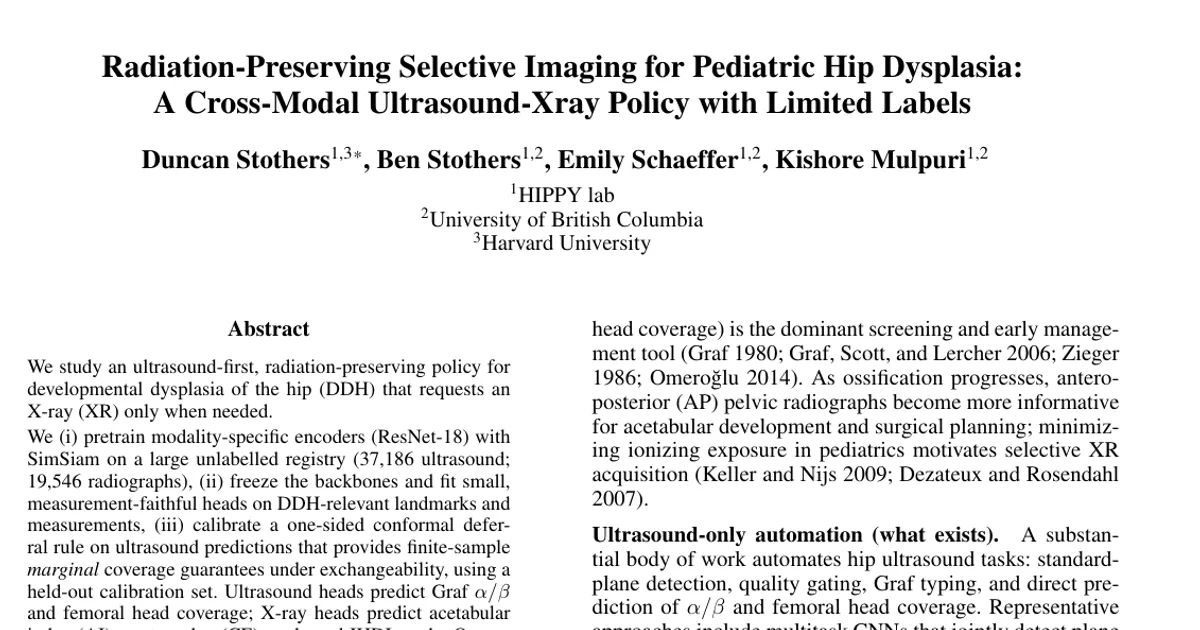
We study an ultrasound-first, radiation-preserving policy for developmental dysplasia of the hip (DDH) that requests a radiograph only when needed. We (i) pretrain modality-specific encoders (ResNet-18) with SimSiam on a large unlabelled registry (37186 ultrasound; 19546 radiographs), (ii) freeze the backbones and fit small, measurement-faithful heads on DDH-relevant landmarks and measurements, (iii) calibrate a one-sided conformal deferral rule on ultrasound predictions that provides finite sample marginal coverage guarantees under exchangeability, using a held-out calibration set. Ultrasound heads predict Graf alpha, beta, and femoral head coverage; X-ray heads predict acetabular index (AI), center-edge (CE) angle and IHDI grade. On our held out labeled evaluation set, ultrasound measurement error is modest (e.g., alpha MAE ~= 9.7 degrees, coverage MAE ~= 14.0%), while radiographic probes achieve AI and CE MAEs of ~= 7.6 degrees and ~= 8.9 degrees, respectively. The calibrated US-only policy is explored across rule families (alpha-only; alpha OR coverage; alpha AND coverage), conformal miscoverage levels, and per-utility trade-offs using decision-curve analysis. Conservative settings yield high coverage with near-zero US-only rates; permissive settings (e.g., alpha OR coverage at larger deltas) achieve non-zero US-only throughput with expected coverage tradeoffs. The result is a simple, reproducible pipeline that turns limited labels into interpretable measurements and tunable selective imaging curves suitable for clinical handoff and future external validation.