Research
PGVMS: A Prompt-Guided Unified Framework for Virtual Multiplex IHC Staining with Pathological Semantic Learning
Overview Research area: Computational pathology and medical image-to-image translation, specifically virtual (AI-generated) immunohistochemical (IHC) staining from hematoxylin and eosin (H&E) slides.
- arXiv
- 2602.23292
- Published
- 2026-02-26
- Authors
- Fuqiang Chen, Ranran Zhang, Wanming Hu, Deboch Eyob Abera, Yue Peng, Boyun Zheng, Yiwen Sun, Jing Cai, Wenjian Qin
AI summary
Overview
Research area: Computational pathology and medical image-to-image translation, specifically virtual (AI-generated) immunohistochemical (IHC) staining from hematoxylin and eosin (H&E) slides.
Technical level: Advanced. The paper assumes familiarity with GANs, contrastive learning (CUT, NCE loss), diffusion-based translation models, stain deconvolution, and pathology vision-language models.
Scope: The paper proposes PGVMS, a single prompt-guided framework that converts an H&E image into multiple IHC stains (ER, PR, HER2, Ki67) using only uniplex training data, and evaluates it on two benchmark datasets (MIST and IHC4BC). The provided content is truncated at the end of the implementation-details section, so the loss-weighting list and any later material are not reported here.
What This Paper Is About
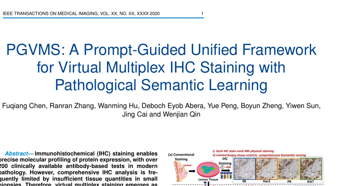
Immunohistochemistry reveals protein biomarkers that H&E staining cannot show, but it requires physical tissue, expensive reagents, and protocols taking up to 60 hours — and small biopsies often do not contain enough tissue for all the antibody tests a pathologist needs. Virtual staining tries to solve this by digitally converting an H&E image into IHC images, but the authors argue that existing methods suffer from three failures: weak semantic control over which stain is generated, inaccurate protein-expression distributions, and spatial misalignment between the H&E input and its IHC label (because the two images come from two separate, depth-wise consecutive tissue cuts). PGVMS is the authors' unified framework for addressing all three at once.
Key Contributions
-
A pathological semantics–style guided (PSSG) generator. It integrates the CONCH pathology vision-language model (trained on 1.17 million pathology image-caption pairs) so that a natural-language prompt such as "H&E to Her2 stained image" controls which stain is produced, with an adaptive image-conditioned bias term that adjusts the prompt embedding to each tumor's morphology. A prompt-guided style normalization (PGSN) module mixes instance normalization and layer normalization with learned, prompt-derived parameters.
-
A protein-aware learning strategy (PALS). It quantifies protein expression directly by computing optical density in the DAB channel (via color deconvolution and the Lambert-Beer law), then applies a novel focal optical density (FOD) map and a multi-level protein awareness (MLPA) loss spanning global intensity, histogram distribution, and regional block consistency.
-
A prototype-consistent learning strategy (PCLS). It extracts protein-expression and normal-tissue prototypes from both generated and real IHC images using a frozen pretrained segmentation U-Net, then enforces bidirectional cross-image prototype consistency to tolerate spatial misalignment between H&E inputs and IHC labels.
-
A unified multiplex staining system. Rather than a separate model per biomarker, PGVMS generates multiple stains in one framework, and the authors report state-of-the-art pathological consistency on the MIST and IHC4BC datasets, while noting experiments on cross-organ clinical datasets as part of the evaluation (results for that dataset are not visible in the provided text).
Main Findings
-
Pathological consistency beats competing methods on MIST. For the four breast-cancer biomarkers, PGVMS reports Pearson-R of 0.8548 (HER2), 0.8902 (ER), 0.9126 (PR), and 0.8018 (Ki67) on the MIST dataset.
-
Strong results on IHC4BC as well. PGVMS reports Pearson-R of 0.7751 (HER2), 0.7940 (ER), 0.7084 (PR), and 0.8913 (Ki67) on the IHC4BC dataset.
-
Not uniformly best on Pearson-R. On MIST Ki67, PSPStain reports a higher Pearson-R (0.8744) than PGVMS (0.8018); PGVMS is highest among all listed methods for HER2, ER, and PR on MIST and for all four markers on IHC4BC.
-
Perception metrics (FID and DISTS) are generally lowest or near-lowest for PGVMS. On MIST, PGVMS FID/DISTS are 44.4247/0.2336 (HER2), 36.5734/0.2299 (ER), 34.9099/0.2300 (PR), 36.3537/0.2439 (Ki67); PSPStain reports a lower FID on MIST HER2 (41.3439) than PGVMS (44.4247). On IHC4BC, PGVMS reports 53.3853/0.2552 (HER2), 36.8306/0.2326 (ER), 40.6377/0.2495 (PR), 24.4450/0.2208 (Ki67).
-
Reference-based quality metrics are not where PGVMS dominates. PGVMS PSNR/SSIM on MIST are 13.9246/0.1769 (HER2), 14.3583/0.2072 (ER), 14.2510/0.2000 (PR), 14.8344/0.2285 (Ki67) — e.g., PyramidP2P reports a higher MIST HER2 PSNR of 14.9122 — while the paper positions its advantage as pathological consistency rather than reconstruction fidelity.
-
One unified model replaces many single-task models. Baseline virtual-staining methods such as PyramidP2P, ASP, TDKStain, and PSPStain are biomarker-specific (one-to-one), whereas PGVMS generates all four stains in a one-to-many setting with prompt control.
-
Diffusion and general-domain prompt baselines underperform on pathology. DDBM shows large FID values (201.5442 for MIST HER2, 191.6672 for MIST ER, 290.9201 for IHC4BC ER), and ControlNet shows low PSNR (8.6754 for MIST HER2, 7.9736 for IHC4BC HER2) with distorted IOD values (up to +25.7725 for IHC4BC ER), which the authors attribute to imprecise histopathological semantic capture.
-
Two hyperparameters were tuned empirically. The focal parameter α = 1.8 gave optimal performance in the authors' experiments, and the global-constraint tolerance was β = 0.2, described as a 20% tolerance threshold; the histogram term used N_h = 20 bins and the regional term used N_b = 16 non-overlapping blocks.
Methodology in Plain English
The system takes an H&E image and a short text prompt naming the stain you want. A pathology-specific vision-language model (CONCH) turns the prompt into an embedding. Because each tumor looks different, the framework also summarizes the H&E image with two pooling branches — average pooling for overall tissue architecture and max pooling for local cellular abnormalities — and adds that summary to the prompt embedding, so the same prompt produces stain characteristics suited to that particular case. A style-normalization block then uses this combined embedding to set the normalization parameters that govern the generated image, blending instance normalization (local staining patterns) with layer normalization (global tissue structure).
Two learning strategies keep the output biologically faithful. The first, PALS, ignores raw pixel color and instead estimates how much protein is present by measuring optical density in the DAB (brown) channel, then applies a focusing exponent (α = 1.8) that suppresses the large normal-tissue background and emphasizes the small protein-expressing regions. Losses then compare generated versus real images at three scales: mean staining intensity (with a 20% tolerance), a 20-bin histogram of intensity distributions spanning weak, moderate, and strong expression, and a 16-block regional comparison that mimics how pathologists assess hotspots.
The second strategy, PCLS, accepts that H&E and IHC images of adjacent tissue cuts will never align pixel-perfectly. Instead of forcing pixel correspondence, a frozen pretrained U-Net segments each image into protein-expression versus normal tissue, computes a confidence-weighted average feature ("prototype") for each class from both the generated and the real image, and then pulls image features toward the opposite image's prototypes in both directions using cosine similarity and softmax. This makes semantically similar content converge even when the spatial positions differ.
Training combines adversarial loss, patch contrastive (NCE) loss, SSIM loss, Gaussian pyramid loss, and the two new strategy losses. The implementation uses PyTorch on an NVIDIA RTX A6000, builds on the CUT framework with a ResNet-6Blocks generator and a PatchGAN discriminator, trains on random 512×512 patches with batch size 1, uses Adam at a fixed learning rate of 1×10⁻⁴ for 80 epochs, and sets λ_M = 1.0, λ_C = 2.5, λ_S = 0.05, λ_G = 10.0 — the provided text truncates at this point.
Why This Matters
Impact on research. The paper reframes virtual multiplex staining as a unified, prompt-controlled generation problem rather than a collection of biomarker-specific models, and it argues that the field's real bottleneck is not pixel-level fidelity but pathology-level semantics — protein expression distribution and cross-modality alignment. It also demonstrates a way to train a multiplex system using only uniplex data, and it introduces measurable mechanisms (optical-density quantification, prototype consistency) that other virtual-staining work could adopt.
Real-world applications:
- Small biopsies from cancer patients, where limited tissue prevents running every needed antibody test, could be digitally re-stained to suggest additional biomarker expression.
- Resource-limited clinical settings without multiplex IHC equipment or the staff time for protocols lasting up to 60 hours could get preliminary multiplex-like views from routine H&E slides.
- Pathology research cohorts with archived H&E slides but no corresponding IHC could be enriched retrospectively with virtual biomarker representations.
- Pathologist training and quality control could use virtual stains to illustrate expected protein patterns across the 200+ clinically available antibody-based tests.
Industry relevance. Digital pathology vendors and AI diagnostics companies are investing heavily in foundation-model-based image analysis; a method that couples a pathology vision-language model (CONCH) to a stain-generation generator points toward a single deployable model serving many stains instead of a per-marker product line. The emphasis on protein-expression quantification also speaks directly to the regulatory and clinical need for outputs that are interpretable in standard pathology terms (0/1+/2+/3+ expression levels) rather than only visually plausible images.
Future Directions
- Closing the gap on markers where it is not best. PGVMS trails PSPStain in Pearson-R on MIST Ki67 (0.8018 versus 0.8744), suggesting that the prototype-consistency strategy may not transfer equally to proliferation markers.
- Reporting the cross-organ clinical evaluation in full. The contributions list a cross-organ clinical dataset as part of the evaluation, but the results are not visible in the provided content, leaving its generalization claim unverified here.
- Separating fidelity from pathology metrics. PGVMS does not lead on PSNR or SSIM, so an open question is whether reference-based reconstruction metrics are the right target for virtual staining, or whether protein-expression metrics should replace them.
- Validating beyond the four breast-cancer biomarkers. All reported experiments use ER, PR, HER2, and Ki67 on MIST and IHC4BC; whether prompt control extends to other markers, tissues, or multi-marker co-staining combinations remains to be shown.
Target Audience
This paper is most useful to computational pathology and medical-imaging researchers working on virtual staining, stain transfer, or foundation-model-guided generation, and to graduate students already comfortable with GANs and contrastive or diffusion-based image translation. Pathologists and clinical informatics teams evaluating whether virtual multiplex staining could substitute for or supplement physical IHC will find the problem framing and the protein-expression metrics relevant, while readers without a machine-learning background will need the methods section explained.
Authors’ abstract
Immunohistochemical (IHC) staining enables precise molecular profiling of protein expression, with over 200 clinically available antibody-based tests in modern pathology. However, comprehensive IHC analysis is frequently limited by insufficient tissue quantities in small biopsies. Therefore, virtual multiplex staining emerges as an innovative solution to digitally transform H&E images into multiple IHC representations, yet current methods still face three critical challenges: (1) inadequate semantic guidance for multi-staining, (2) inconsistent distribution of immunochemistry staining, and (3) spatial misalignment across different stain modalities. To overcome these limitations, we present a prompt-guided framework for virtual multiplex IHC staining using only uniplex training data (PGVMS). Our framework introduces three key innovations corresponding to each challenge: First, an adaptive prompt guidance mechanism employing a pathological visual language model dynamically adjusts staining prompts to resolve semantic guidance limitations (Challenge 1). Second, our protein-aware learning strategy (PALS) maintains precise protein expression patterns by direct quantification and constraint of protein distributions (Challenge 2). Third, the prototype-consistent learning strategy (PCLS) establishes cross-image semantic interaction to correct spatial misalignments (Challenge 3).