Research
Assessing nnU-Net Generalization across Brain Tumor Populations in BraTS-GoAT 2026
Overview Research area: Medical image analysis — 3D brain tumor segmentation with deep learning, evaluated in the BraTS-GoAT 2026 benchmark setting. Technical level: Advanced. The work assumes familia
- arXiv
- 2609.15524
- Published
- 2026-09-14
- Authors
- Tristan Kirscher, Vivian Metzger, Philippe Meyer, Xavier Coubez
AI summary
Overview
Research area: Medical image analysis — 3D brain tumor segmentation with deep learning, evaluated in the BraTS-GoAT 2026 benchmark setting.
Technical level: Advanced. The work assumes familiarity with nnU-Net, cross-validation, Dice similarity coefficient (DSC), test-time augmentation, and volume-adjusted statistical analysis.
Scope: A single paper assessing how well a standard 3D nnU-Net segmentation model generalizes from the population it was trained on to a more heterogeneous pooled validation population, and characterizing which cases fail.
What This Paper Is About
Segmentation models for brain tumors are usually trained and validated on cases drawn from a limited set of sources, which raises the question of whether they still work when applied to a broader, more varied patient population. BraTS-GoAT is a benchmark designed to test exactly that, evaluating tumor segmentation across heterogeneous populations. The goal of this paper is to measure, rather than assume, how much performance a conventional nnU-Net loses when it moves from source out-of-fold cases to pooled official validation, and to explain what distinguishes the cases it gets wrong.
Key Contributions
- A conventional nnU-Net baseline for BraTS-GoAT. A standard 3D nnU-Net was trained on 1,351 labeled cases using five-fold cross-validation with 1,000 epochs per fold.
- An explicit generalization-gap measurement. The authors compared performance under matched fold-0 inference between source out-of-fold cases and pooled official validation, isolating the drop attributable to the population shift.
- An evaluation of inexpensive inference tricks. The final predictor averaged all folds and applied test-time mirroring; the paper reports what mirroring and fold ensembling actually contributed, and also tests a residual-encoder alternative.
- A failure-mode analysis with volume adjustment. Rather than only reporting failure by raw tumor size, the authors adjusted for enhancing tumor (ET) and whole tumor (WT) volume to identify structural properties of the ET segment that remain associated with lower Dice.
Main Findings
- Pooled validation scores: On pooled official validation, global DSC was 0.7805 for enhancing tumor (ET), 0.8288 for tumor core (TC), and 0.8854 for whole tumor (WT) — ET is the hardest of the three regions.
- A measurable generalization gap: Under matched fold-0 inference, mean regional Dice fell from 0.9058 on source out-of-fold cases to 0.8310 on pooled validation, a difference of −0.0747.
- Mirroring did not deliver as an ensemble: Test-time mirroring produced small gains within a single fold, but gave no clear benefit in the ensemble setting.
- A residual-encoder alternative did not beat the baseline: The residual-encoder variant reached 0.8282 mean Dice, below the reported matched fold-0 baseline figure and in the same range as the CNN-style result.
- Failures concentrate in small enhancing tumors: Among labeled out-of-fold predictions, failure cases had substantially smaller reference ET volumes.
- Size alone does not explain failures: After adjustment for ET and WT volume, lower Dice remained associated with a more disconnected ET (more separate components) and with a smaller fraction of the ET falling inside its largest component.
Methodology in Plain English
The team trained the most standard, well-established 3D segmentation pipeline available — nnU-Net in its conventional configuration — on 1,351 labeled cases, splitting them into five folds so that each case served as unseen data once, and training each fold for 1,000 epochs. The predictor used for submission averaged the outputs of all five folds and also mirrored the input at test time (a common trick that feeds a flipped copy of the image and combines the two predictions). To separate "how good is the model" from "how much does the new population hurt," the authors ran a matched comparison restricted to fold 0, evaluating it both on its own held-out source cases and on the pooled validation set. They also tried swapping in a residual-encoder variant as an architectural alternative, and then examined which individual cases failed, testing whether failure was explained by tumor volume alone or by additional properties of the enhancing tumor's shape — specifically how fragmented the enhancing region was.
Why This Matters
Research impact. The paper reframes benchmark performance as a generalization question. Reporting a 0.9058 in-distribution score alongside a 0.8310 pooled score makes the population shift a first-class number rather than a caveat, and the volume-adjusted failure analysis points to fragmentation of the enhancing tumor as a difficulty signal that size alone would hide. The negative results — no clear ensembling benefit from mirroring, and a residual encoder that did not improve on the conventional design — are useful counterweights to the assumption that architectural or augmentation changes automatically help.
Real-world applications (potential):
- Multi-site and multi-scanner clinical deployment, where the "population" at inference differs from the training cohort.
- Longitudinal tumor measurement and treatment-response assessment, where ET, TC, and WT must be separated reliably and small enhancing components matter.
- Case triage and quality control, using the identified failure signatures (small, fragmented enhancing tumor) to flag scans for human review.
- External validation studies and regulatory submissions, where a quantified generalization gap supports more honest performance claims.
Industry relevance. Developers of medical imaging AI need to know how much accuracy is lost outside their training distribution and where to invest engineering effort. The findings suggest that gains may lie in handling fragmented, low-volume enhancing tumor rather than in simply scaling epochs, swapping encoders, or adding fold ensembling.
Future Directions
- Target the specific failure mode. Methods that preserve connectivity of small, fragmented enhancing tumor — such as topology-aware losses or component-level post-processing — are the natural next step given that disconnected ET remained associated with lower Dice after volume adjustment.
- Test stronger generalization strategies. The abstract evaluates mirroring and a residual encoder but not explicit domain adaptation or cross-population training; whether those close the −0.0747 gap is unresolved.
- Re-examine inference-time ensembling. Since mirroring helped within a fold but not in the ensemble, the interaction between test-time augmentation and multi-fold averaging deserves a more systematic study.
- Extend the volume-adjusted analysis. The abstract reports an association, not a mechanism; understanding why fragmented enhancing tumor is harder, and whether it holds beyond out-of-fold labeled cases, remains open.
Target Audience
Researchers and engineers working on medical image segmentation who want a rigorous, sober assessment of out-of-distribution generalization rather than a leaderboard score. It is also relevant to clinicians and imaging scientists evaluating whether automated tumor segmentation is trustworthy across varied patient populations, and to benchmark organizers and reviewers interested in how to report in-distribution versus pooled performance side by side. The paper assumes comfort with nnU-Net, cross-validation, and Dice-based metrics, so beginners may need background reading first. Details that the abstract does not provide — full per-region breakdowns, statistical tests, and the complete comparison of the residual-encoder variant — would require the full text.
Authors’ abstract
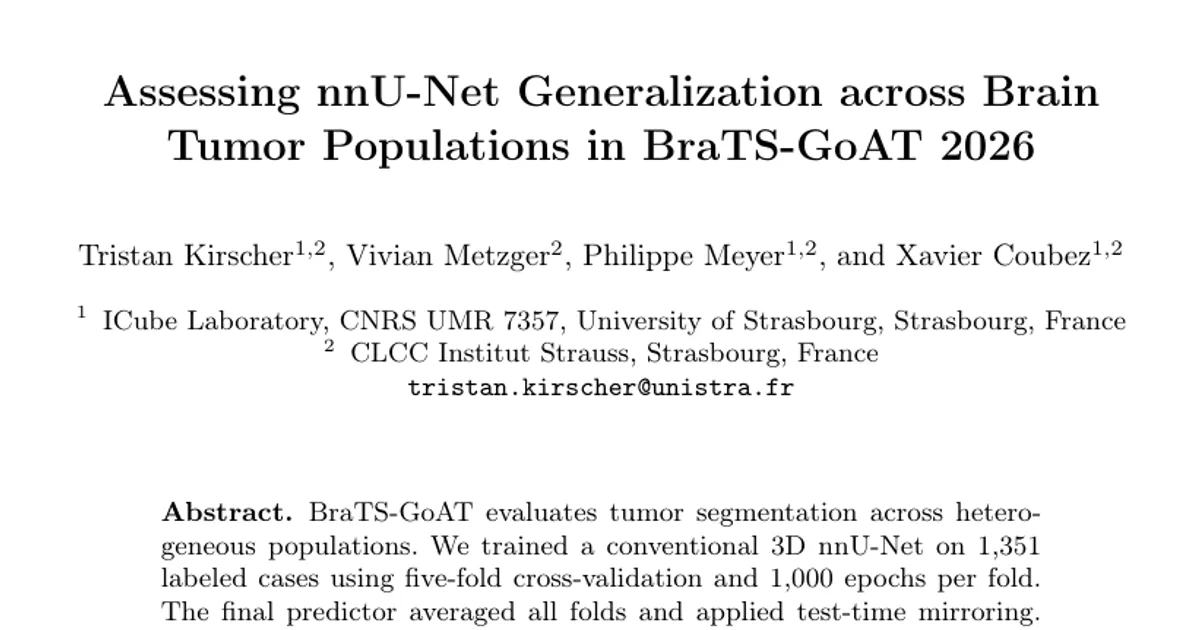
BraTS-GoAT evaluates tumor segmentation across heterogeneous populations. We trained a conventional 3D nnU-Net on 1,351 labeled cases using five-fold cross-validation and 1,000 epochs per fold. The final predictor averaged all folds and applied test-time mirroring. On pooled official validation, global DSC values were 0.7805, 0.8288, and 0.8854 for enhancing tumor (ET), tumor core (TC), and whole tumor (WT). Under matched fold-0 inference, mean regional Dice decreased from 0.9058 on source out-of-fold (OOF) cases to 0.8310 on pooled validation (difference--0.0747). Mirroring gave small single-fold gains but no clear ensemble benefit; a residual-encoder alternative reached 0.8282 mean Dice. In labeled OOF predictions, failure cases had substantially smaller reference ET volumes; after adjustment for ET and WT volume, lower Dice remained associated with more disconnected ET components and a smaller fraction of ET contained in the largest component.